Ossification
This is the process by which the
mesenchymal cells and the cartilages are converted into bones during
development. Initially, during embryonic development, the skeleton remains
primarily cartilaginous to form the basic structural components and framework
of the body. Thereafter when the basic structure of the embryo is
formed, bone begins to be deposited. There are two ways in which bones are
deposited;
Intramembranous ossification: Is the process of bone formation in which the mesenchyme differentiated directly into the bone, example, is the flat bones of the skull.
In this process, the mesenchyme first differentiates in to osteoblasts (bone–forming cell) which then begins to deposits osteoid (unmineralized matrix). Thereafter the osteoblasts deposit calcium phosphate into the osteoid tissue, and the osteoid then is converted into bone.
The osteoblasts are transformed and become the osteocyts. Initially, the developing bone has no organized pattern. Soon, the spicules (fig. 8) of the bones becomes organized and coalesce into lamellar (layers). Different lamellar develop around blood vessels forming osteon (Haversian canal system). Other osteoblasts remain at the surface of the developing bone and continue to lay down lamellae forming the compact bone. While between the surface plates, the intervening bone remains spongy. Similarly the mesenchyme differentiates into bone marrow.
Note that: During fetal and postnatal life, there is continuous remodeling of bone by the coordinated action of osteoclasts and osteoblasts.
Endochondral ossification: This is the process of bone formation in which the mesenchymal cells give rise to cartilaginous models first which in turn become ossified and form bone (fig. 4 A-E). The cartilage is gradually replaced by bones, examples are long bones of the limbs, basal bones of the skull, vertebral column and ribs.
This process involves cells hypertrophy, deposition of calcium phosphate crystals, cell death and erosion of calcium matrix (fig. 4 C-H). Concurrently, a thin layer of bone is deposited under the perichondrium (covering of the cartilage), thus the perichondrium becomes the periosteum (outer layer of the bone)
There are two centers of ossification, the primary and the secondary. The primary centers of ossification appear in the diaphysis, and the process continues toward the epiphysis (fig. 4C) as in the long bone while the secondary centers appear in the epiphysis in most bones particularly during the first few years after birth (fig.4H). At birth, the diaphyses are largely ossified but most of the epiphyses are still cartilaginous
Ossification spreads radially, and only the articular cartilage, a transverse plate of cartilage and the epiphysial cartilage plate, remain cartilaginous
- Intramembranous ossification
- Endochondral ossification
Intramembranous ossification: Is the process of bone formation in which the mesenchyme differentiated directly into the bone, example, is the flat bones of the skull.
In this process, the mesenchyme first differentiates in to osteoblasts (bone–forming cell) which then begins to deposits osteoid (unmineralized matrix). Thereafter the osteoblasts deposit calcium phosphate into the osteoid tissue, and the osteoid then is converted into bone.
The osteoblasts are transformed and become the osteocyts. Initially, the developing bone has no organized pattern. Soon, the spicules (fig. 8) of the bones becomes organized and coalesce into lamellar (layers). Different lamellar develop around blood vessels forming osteon (Haversian canal system). Other osteoblasts remain at the surface of the developing bone and continue to lay down lamellae forming the compact bone. While between the surface plates, the intervening bone remains spongy. Similarly the mesenchyme differentiates into bone marrow.
Note that: During fetal and postnatal life, there is continuous remodeling of bone by the coordinated action of osteoclasts and osteoblasts.
Endochondral ossification: This is the process of bone formation in which the mesenchymal cells give rise to cartilaginous models first which in turn become ossified and form bone (fig. 4 A-E). The cartilage is gradually replaced by bones, examples are long bones of the limbs, basal bones of the skull, vertebral column and ribs.
This process involves cells hypertrophy, deposition of calcium phosphate crystals, cell death and erosion of calcium matrix (fig. 4 C-H). Concurrently, a thin layer of bone is deposited under the perichondrium (covering of the cartilage), thus the perichondrium becomes the periosteum (outer layer of the bone)
There are two centers of ossification, the primary and the secondary. The primary centers of ossification appear in the diaphysis, and the process continues toward the epiphysis (fig. 4C) as in the long bone while the secondary centers appear in the epiphysis in most bones particularly during the first few years after birth (fig.4H). At birth, the diaphyses are largely ossified but most of the epiphyses are still cartilaginous
Ossification spreads radially, and only the articular cartilage, a transverse plate of cartilage and the epiphysial cartilage plate, remain cartilaginous
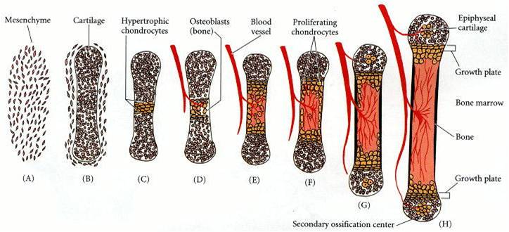
Fig. 4 (Copyright © 2006 Sinuer Associates), Different stages of endochondral ossification in long bone (Gilbert, 2006)
Growth and lengthening of long bone

Fig. 5 (Copyright © 2001 Addison Wesley),
The diaphysial-epiphysial
junction
is the spot where lengthening in the long bone occur. The chondrocytes of the epiphysial cartilage plates (growth plates) (fig. 4H) proliferate (by mitosis)
and participate in endochondral bone formation.
Growth in the diameter of a bone results from deposition of bone at the periosteum and from resorption on the internal medullary surface (fig 6). The rate of deposition and resorption is balanced to regulate the thickness of the compact bone and the size of the medullary cavity (fig. 5)
Growth in the diameter of a bone results from deposition of bone at the periosteum and from resorption on the internal medullary surface (fig 6). The rate of deposition and resorption is balanced to regulate the thickness of the compact bone and the size of the medullary cavity (fig. 5)

Fig. 6 (Copyright © 2004 McGraw-Hill), (Seeley et al., 2004)
**********************************************************************************************************************************
(Copyright © 2011 by U. Bala)
