Embryological Source of Skeletal System
The skeletal system develops broadly from
two main sources, the mesoderm and the ectoderm. Specifically, from the
mesoderm are the paraxial and lateral (somatic) plate mesoderm and from the
ectoderm is the neural crest cells.
The Paraxial mesoderm forms somites (fig.2 A); which are segmented series of tissue blocks on each side of the neural tube.The somites differentiate into;
The sclerotome cells differentiate and form the mesenchyme by the end of the 4th week. The mesenchyme consists of loosely packed, unspecialised cells set in a gelatinous ground substance. One of the main characteristic of mesenchymal cells is that, they migrate and differentiate in many ways; they may become fibroblasts, chondroblasts or osteoblasts which will later gives rise to connective tissues, cartilages and the bones respectively.
Thelateral plate mesoderm differentiates into in to mesenchymal cells that contribute mesodermal cells for the formation of the pelvic and shoulder girdles and the long bones of the limbs.
The Paraxial mesoderm forms somites (fig.2 A); which are segmented series of tissue blocks on each side of the neural tube.The somites differentiate into;
- Sclerotome; the cells from this region form the bony and cartilaginous component of the body, mainly the vertebrae and the ribs (fig 2 A-D)
- Dermomyotome; the cells
from its myotome region form myoblasts (primordial muscle cells), and those
from its dermatome region form the dermis (fibroblasts) (fig 2A-D).
The sclerotome cells differentiate and form the mesenchyme by the end of the 4th week. The mesenchyme consists of loosely packed, unspecialised cells set in a gelatinous ground substance. One of the main characteristic of mesenchymal cells is that, they migrate and differentiate in many ways; they may become fibroblasts, chondroblasts or osteoblasts which will later gives rise to connective tissues, cartilages and the bones respectively.
Thelateral plate mesoderm differentiates into in to mesenchymal cells that contribute mesodermal cells for the formation of the pelvic and shoulder girdles and the long bones of the limbs.
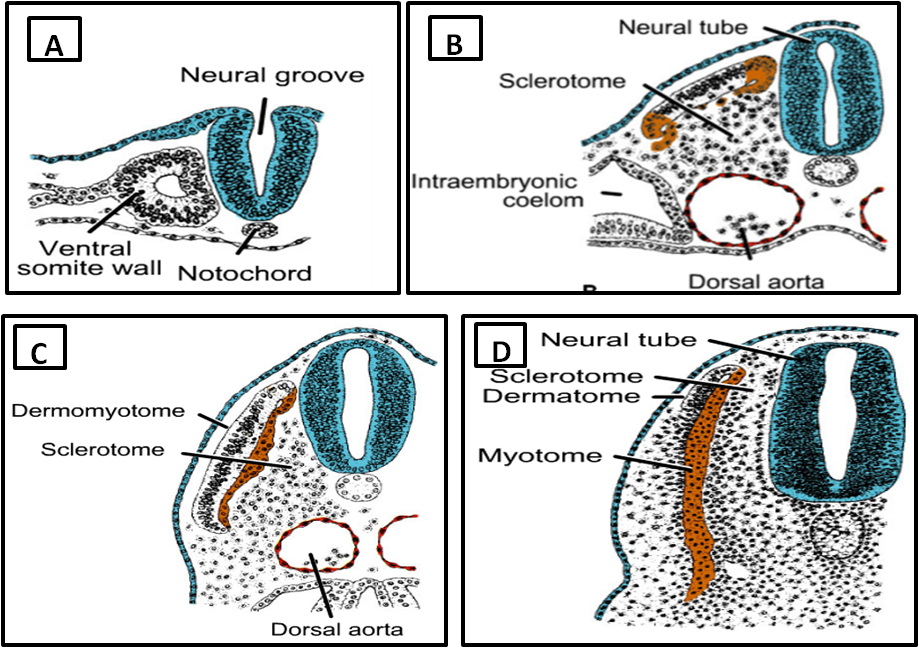
Fig. 2 (Copyright © 2010 Wolters Kluwer); Development of somites; A. Paraxial mesoderm; B & C. Ventromedial part (sclerotomes) and dorsolateral part (dermomyotome) of paraxial mesoderm. D Further differentiation of the dermomyotome in to myotome and dermatome (Sadler, 2010)
Neural crest cells
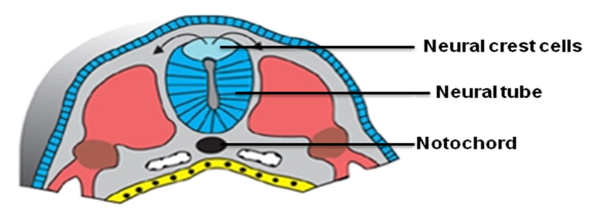
Fig. 3 (Copyright © 2010 Wolters Kluwer) (Sadler, 2010)
The neural crest cells
(fig. 3) formed at the tip of the neural tube and they do not migrate from this region until the neural tube closure is complete. These cells differentiate into mesenchyme which participate
in the formation of bones of the face and skull.
**********************************************************************************************************************************
(Copyright © 2011 by U. Bala)
