The vertebral column
A vertebra consist of different parts which originate from the the sclerotomal cells. Early during the fourth week of development these sclerotomal cells from the somites surround the;
Those cells ventromedial to the notochord arrange themselves in alternating bands of loose and densely packed cells (fig. 13 A). The centrum of the vertebra is formed when a region of the packed cells fuses with a region of loose cells immediately caudal which has arisen from an adjacent sclerotome. Some densely packed cells from each region also migrate cranially opposite the myotome to form the intervertebral disc. The intersegmented nerves lie close to the discs while the arteries lie close to the vertebral bodies (fig. 13 B & C).
The notochord degenerates and disappears. It persists only between the vertebrae as the nucleus pulposus. This nucleus is later surrounded by circularly arranged fibers that form the anulus fibrosus (fig. 13B). The nucleus pulposus and anulus fibrosus together make up the intervertebral disc. The neural arch is form from the mesenchymal cells surrounding the neural tube while those cells in the body wall form the costal processes that form ribs in the thoracic region.
Chondrification begins in week six. Ossification begins before birth and end during the 25th year. At birth, three primary ossification centers are present in the centrum and in each half of the vertebral (neural) arch
Note that:
A vertebra consist of different parts which originate from the the sclerotomal cells. Early during the fourth week of development these sclerotomal cells from the somites surround the;
- Ventromedial aspect of the notochord to form the centrum and the intervertebral disc
- Dorsal portion of the neural tube to form the neural arch, and
- Ventrolateral aspect of the body wall to form the costal processes.
Those cells ventromedial to the notochord arrange themselves in alternating bands of loose and densely packed cells (fig. 13 A). The centrum of the vertebra is formed when a region of the packed cells fuses with a region of loose cells immediately caudal which has arisen from an adjacent sclerotome. Some densely packed cells from each region also migrate cranially opposite the myotome to form the intervertebral disc. The intersegmented nerves lie close to the discs while the arteries lie close to the vertebral bodies (fig. 13 B & C).
The notochord degenerates and disappears. It persists only between the vertebrae as the nucleus pulposus. This nucleus is later surrounded by circularly arranged fibers that form the anulus fibrosus (fig. 13B). The nucleus pulposus and anulus fibrosus together make up the intervertebral disc. The neural arch is form from the mesenchymal cells surrounding the neural tube while those cells in the body wall form the costal processes that form ribs in the thoracic region.
Chondrification begins in week six. Ossification begins before birth and end during the 25th year. At birth, three primary ossification centers are present in the centrum and in each half of the vertebral (neural) arch
Note that:
- The two halves of the vertebral arch fuse during the 3-5 years
- The spinal cord enlarge due to the present of the cartilaginous joint between the arch and the centrum. This joint disappear between the 3-6 years after the arch fuses with the centrum.
- After birth, the thoracic vertebral column gradually develops a relatively fixed curve.
- When the infant begins to lift to lift it, the cervical curves appears
- hen the infant begins to walk the
lumber curve appears, usually at the end of the first year
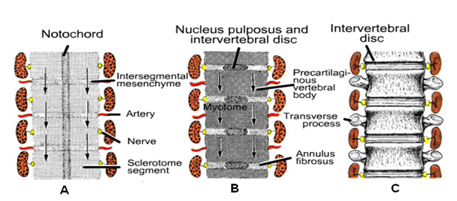
Fig. 13 (Copyright © 2010 Wolters Kluwer); Various stages of the development of vertebral column; A. Sclerotomic segments are separated by less dense intersegmental tissue, B. Condensation and proliferation of the sclerotomal cells around the notochord, C. The vertebral body forms from the cranial and caudal halves of the two successive sclerotomal masses (Sadler, 2010)
**********************************************************************************************************************************
(Copyright © 2011 by U. Bala)
