Development of the Skull
The skull (cranium) (fig.7) develops from mesenchyme around the developing brain. Basically, the cranium consists of two parts:
Neurocranium covers the ventral, lateral and posterior parts of the brain as well as the ear and nose. It is divided into two portions;
The membranous neurocranium as the name implies is formed by intramembranous ossification. The mesenchymal cells are derived from neural crest and paraxial mesoderm. These cells then encircle the brain and form most of the flat bones of the skull. These flat bones are characterized by the present of bone spicules (fig. 8). The spicules progressively spread out from primary ossification centers toward the surface. The flat bones of the skull,like the frontal, parietals, the squamous part of the temporal, the occipital, the nasal and lacrimal bones are all membranous.
The skull (cranium) (fig.7) develops from mesenchyme around the developing brain. Basically, the cranium consists of two parts:
- The Neurocranium; a protective case for the brain
- The Viscerocranium; the skeleton of the face,
Neurocranium covers the ventral, lateral and posterior parts of the brain as well as the ear and nose. It is divided into two portions;
- The membranous neurocranium
- The cartilaginous neurocranium (chondrocranium)
The membranous neurocranium as the name implies is formed by intramembranous ossification. The mesenchymal cells are derived from neural crest and paraxial mesoderm. These cells then encircle the brain and form most of the flat bones of the skull. These flat bones are characterized by the present of bone spicules (fig. 8). The spicules progressively spread out from primary ossification centers toward the surface. The flat bones of the skull,like the frontal, parietals, the squamous part of the temporal, the occipital, the nasal and lacrimal bones are all membranous.

Fig. 7 (Copyright © 2010 Wolters Kluwer); Skeletal structure of skull; mesenchyme for these structures is derived from neural cells (blue), lateral plate mesoderm (yellow) and paraxial mesoderm (somites) (red), (Sadler, 2010)

Fig. 8 (Copyright © 2010 Wolters Kluwer); Flat bones of the skull of 3 month old fetus showing the spread of spicules from the primary ossification center, (Sadler, 2010)
New born skull
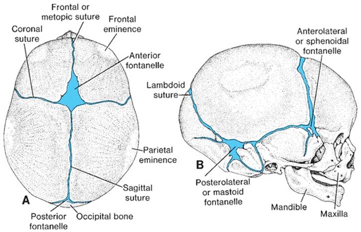
Fig. 9 (Copyright © 2010 Wolters Kluwer) (Sadler, 2010)
The plates of the membranous bones
making up the calvarium of the skull are each derived from the primary ossification center, from which bone formation spreads outward. However, the
individual plates do not fused with each other during prenatal development. As
a consequence, new born babies have unclosed sutures and fontanelles (fig 9).
These temporary discontinuities between the bones of the calvarium aid passage
of the head through the birth canal at childbirth and permit an increase in the
size of the skull to match brain growth after birth. The posterior (smaller)
fontanelle closes during the first year, and the anterior (larger) fontanelle
closes during the second year after birth. However some of the sutures remain
open until adulthood.
Chondrocranium
The chondrocranium is formed by a combination of mesodermal sclerotome and neural crest cells. During development, cartilage are form around the brain beginning at the notochord. Therefore with reference to sella turcica, bones that lie rostral to this point are derived from neural crest cells while the paraxial mesoderm give rise to bones that lie posterior to this limit. Thus, the base of the skull is form when the cartilages formed from these two source fuse and ossify by endochondral ossification (fig. 10).
The parachordal cartilage and the occipital sclerotomes fused to form the base of occipital bone, while the sphenoid and ethmoidal bones are formed from the hypophysial cartilage and the trabeculae cranii respectively. These two bones (sphenoid and ethmoid) lie rostral to occipital bone.
The lesser and greater wing of the sphenoid bone are derived from mesenchymal condensation called the ala orbitalis and ala temporalis respectively. These alae arise by the side of the body of the sphenoid bone. While the petrous and mastoid parts of the temporal bone are derived from periotic capsule. Finally, all these pieces of bones fuse with each other to form a strong base of the skull, expect for the openings via which the cranial nerves leaves the skull (fig. 10)
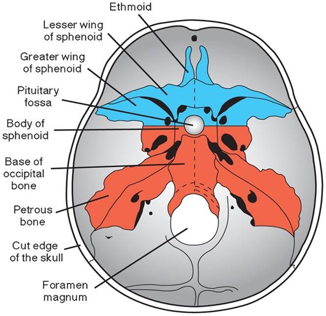
Fig. 10 (Copyright © 2010 Wolters Kluwer); Base of the skull in adult showing the various embryonic component participate in its formation. Part derived from neural crest cells (blue) and paraxial mesoderm (red). (Sadler, 2010)
Viscerocranium
This consists of the bones of the face. They are formed mainly from the first two pharyngeal arches, they are also divided in to membranous viscerocranium and chondral viscerocranium.
Membranous Viscerocranium
The first pharyngeal arch has the dorsal and ventral portion (fig 11). The dorsal portion (maxillary process) undergoes intramembranous ossification and gives rise to the maxilla, the zygomatic bone, the squamous temporal bones, the vomer and the palatine bone, but the squamous temporal bones later become part of the neurocranium. The ventral portion (mandibular process) contains the Meckel’s cartilage (fig 11) and this region become surrounded by mesenchymal cells that condenses and ossifies by membranous ossification to form the mandible. But mandibular condyle undergoes endochondral ossification.
Chondral Viscerocranium
Generally, the neural crest cells give rise to mesenchyme in the head region.These cells migrate into the pharyngeal arches and form the bones and connective tissue of face. The cartilaginous skeleton of the first two pairs of the pharyngeal arch develop in to these part of the fetal cranium.
Cartilage at the dorsal end of the first arch (Meckel’s cartilage) forms the malleus and incus while cartilage at the dorsal end of the second arch (Reichert’s cartilage) forms the stapes and the styloid process (fig. 12). Ossification of the three ossicles begins in the fourth month, making these the first bones to become fully ossified. The ventral end of the second arch ossifies and forms the lesser cornu and the upper body of the hyoid bone. Arch three (ventral end) forms the greater cornu and lower body of the hyoid bone (fig.12)
Finally, arches four and six fuse to form the laryngeal cartilages with the exception of the epiglottis (formed from the hypobrachial eminence of the arches three and four) (fig. 13). In arches three to six, cartilage bears only form at the ventral end of the arch.
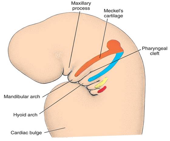
Fig. 11 (Copyright © 2010 Wolters Kluwer); Lateral view of the head and neck showing the cartilages of pharyngeal arches participating in the formation of bones of the face and neck (Sadler, 2010)
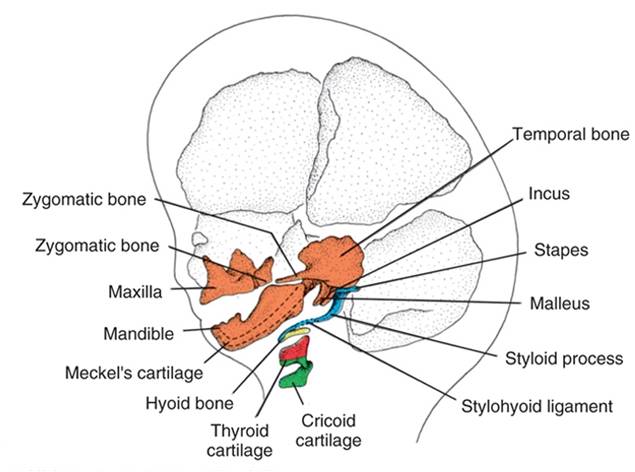
Fig. 12 (Copyright © 2010 Wolters Kluwer); Bones derived from the arch cartilages that contribute to the formation of the face, 1st arch (orange), 2nd arch (blue), 3rd arch (yellow) and 4th arch (red) and 6th arch (green) (Sadler, 2010)
**********************************************************************************************************************************
(Copyright © 2011 by U. Bala)
