Development of Appendicular Skeleton
Limbs are derived from the somatic layer of lateral mesoderm. The mesenchymal cells of this region become activated and the limb buds become visible as an outpocketing from the ventrolateral body wall near the end of the fourth week of development and undergoes a dramatic morphological changes by the end of the embryonic period (fig. 16)
Early during development, the mesenchyme destined for the limbs is covered by a layer of ectoderm.This ectoderm thickens and forms the epical ectodermal ridge (AER) (fig. 15) which exerts an inductive influence on the limb mesenchyme that initiates growth and development of the limbs in a proximodistal axis.
The mesenchyme adjacent to the AER consists of undifferentiated, rapidly proliferating cells, whereas mesenchymal cells proximal to it differentiate into blood vessels and cartilage-bone models. Basically, the bones and the connective tissues of the limbs develop (by endochondral ossification) from this mesenchyme.
The distal end of the limb buds become flattened to form the handplates and footplates and are separated from the proximal segment of the limbs by a circular constriction. Another constriction later appear and further divides the proximal segment in to two segments.
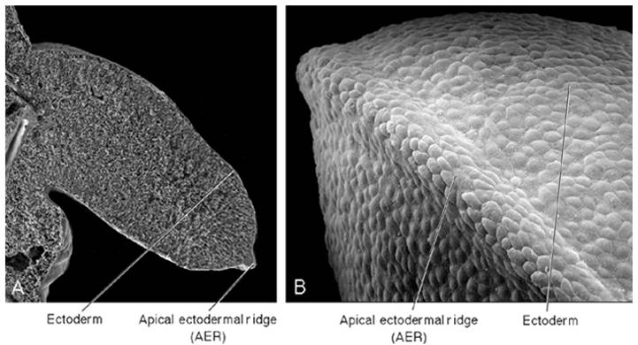
Fig. 15 (Copyright © 2010 Wolters Kluwer); Longitudinal section through the limb bud (A) showing the core mesenchyme covered by a layer of ectoderm, which thickens to form apical ectodermal ridge (Sadler, 2010)
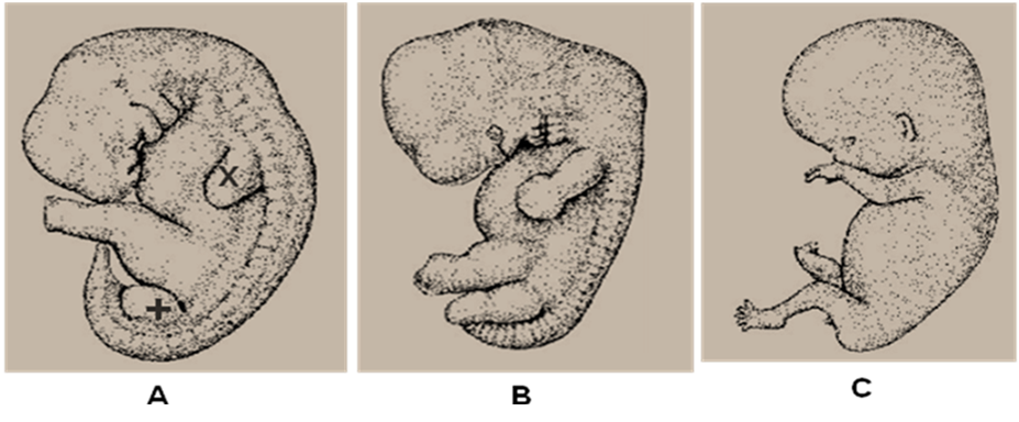
Fig. 16 (Copyright © 2010 Wolters Kluwer); Various stages of limb bud development in human embryo; A. Appearance of the limb buds upper (x) and lower (+) limbs at the end of week four, B. Elongation of the limbs and formation of constrictions at week six, C. Fully formed limb at week eight (Sadler, 2010)
Process of fingers and toes formation

Fig. 17 Zones of cell death during digits formation
The fingers and toes are formed when the mesenchyme of the handplates and the footplates condensed to form digital rays by apoptosis (programme cell death) (fig. 17A-D). Further
formation of the digits depends on;
- Formation of the cartilaginous digital rays by condensation of the mesenchyme
- The death (by apoptosis) of the intervening tissue between the rays
Similarly, as the shape of the limbs is being formed, mesenchyme in the buds condenses and differentiates into
chondrocytes. These cells produced the first hyaline cartilage models foreshadowing the bones of the extremities (fig. 18A) and the entire limb
skeleton is cartilaginous by the end of the sixth week of development (fig.
18B)
Joints are formed when chondrogenesis is arrested and a joint interzone is induced and subsequently joints are form. Joint cavity is formed by apoptosis of the proliferated cells in this zone, while the surrounding cells differentiate in to joint capsule
Development of the upper and lower limbs is similar, except that, the upper limb appeared approximately 1 or 2 days ahead of the lower limb. The upper limb buds develop opposite the cervical segments and the lower limb buds form opposite the lumbar and upper sacral segments (fig. 16 A). Because of their form and function, there are many distinct differences between the development of the hand and foot as in case of rotation of the limb
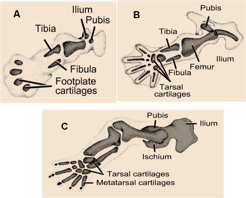
Fig. 18 (Copyright © 2010 Wolters Kluwer); A. The first hyaline cartilage models formed; Complete set of the cartilage models at the end of 6th week (B) and the beginning of 8th week (C) (Sadler, 2010)
Rotation of the limbs
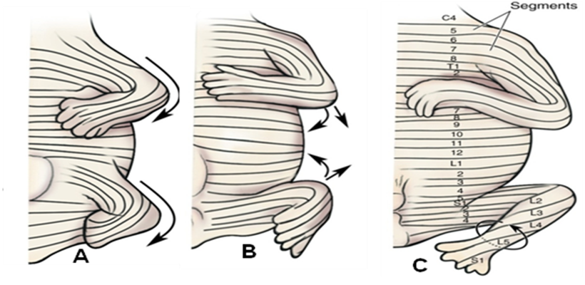
Fig. 19 (Copyright © 2010 Wolters Kluwer) (Moore et al., 2010)
The developing limb rotates in
opposite directions during the seventh week of gestation (fig.19)
The upper limb rotates 90° laterally, so that;
The lower limb rotates approximately 90° medially, placing
The upper limb rotates 90° laterally, so that;
- The extensor muscles is positioned
on the posterior surface,
- The future elbows place dorsally, and
- The thumb is positioned laterally,
The lower limb rotates approximately 90° medially, placing
- The extensor muscles is positioned on the anterior surface,
- The future knees place ventrally and
- The big toe is positioned medially.
By the end of the embryonic period,
primary ossification begins in the diaphysis of the long bones and endochondoral
ossification gradually progresses from diaphysis of the bone
toward
the end of the cartilaginous model (fig 20 arrow).
The (shaft) diaphysis of the long bone is fully ossified at birth, but the epiphysis is still cartilaginous and secondary ossification centers appear in the epiphyses of these bones (fig 20).
The epiphysial plates (growth plate) is temporarily formed between the diaphysial and epiphysial ossification centers.
(fig 20). The persistence of the growth plates provide for interstitial growth in the length of the long bone, whereas periostuem provides for appositional growth in the girth of these bones. Afterwards endochondral ossification advances on both sides of the plate and finally the plate disappear and the epiphysis unite with the shaft of the bone when bone has acquired its full length.
Note that:
- Ossification of the carpal (wrist) bones only begins during the first year after birth
- Developmentally, the radius and the tibia are similar bones, as are the ulna and fibula, just as the thumb and great toe are similar digits
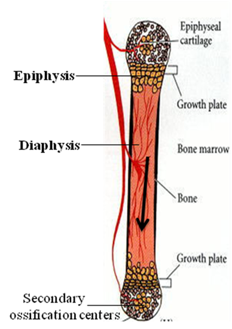
Fig. 20 (Copyright © 2006 Sinuer Associates); Ossification in long bone showing the primary centers (diaphysis) and the direction toward the secondary centers (epiphysis). (Gilbert, 2006)
**********************************************************************************************************************************
(Copyright © 2011 by U. Bala)
